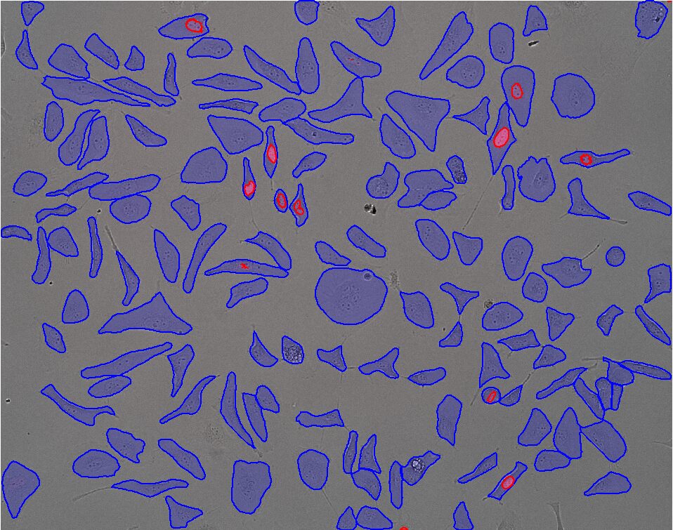
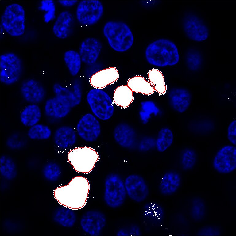
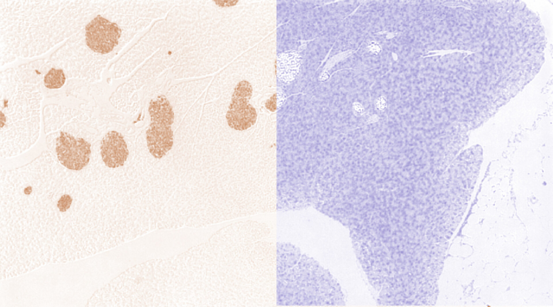


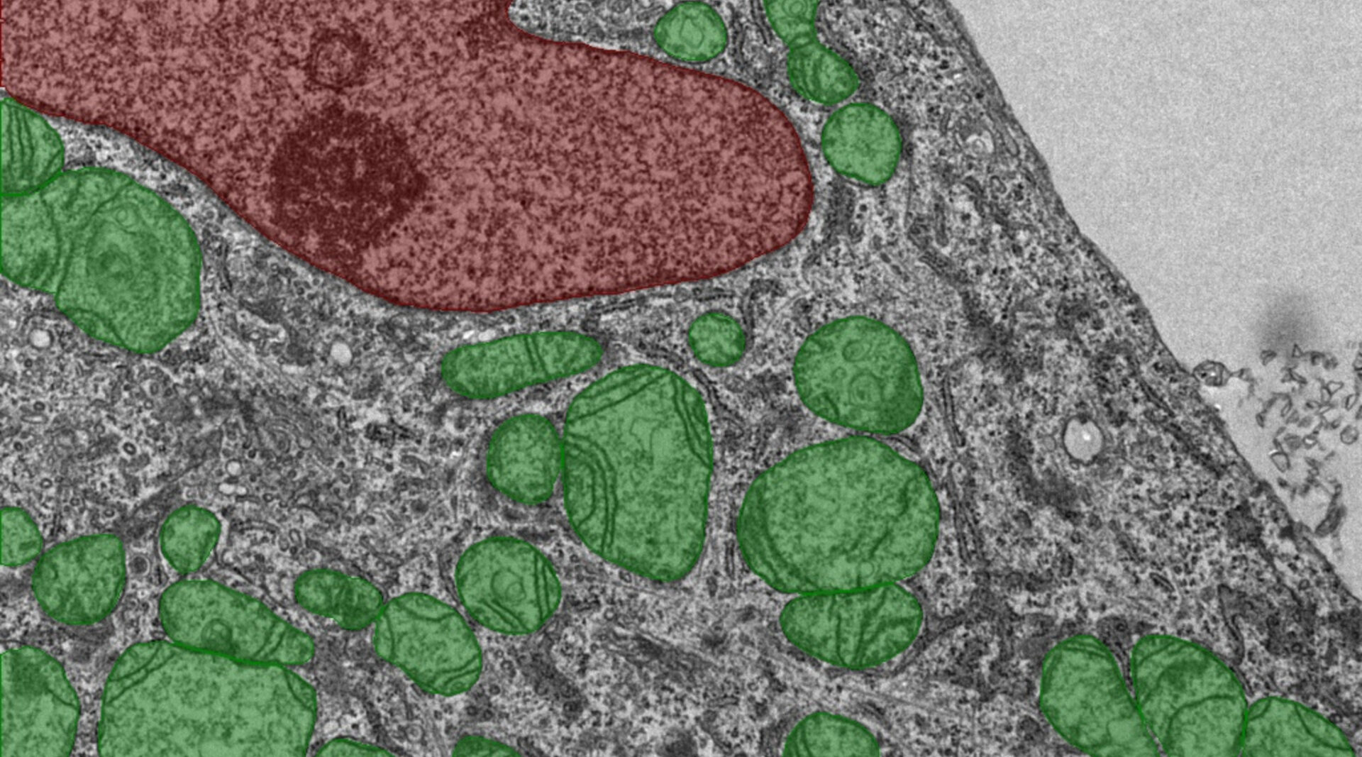
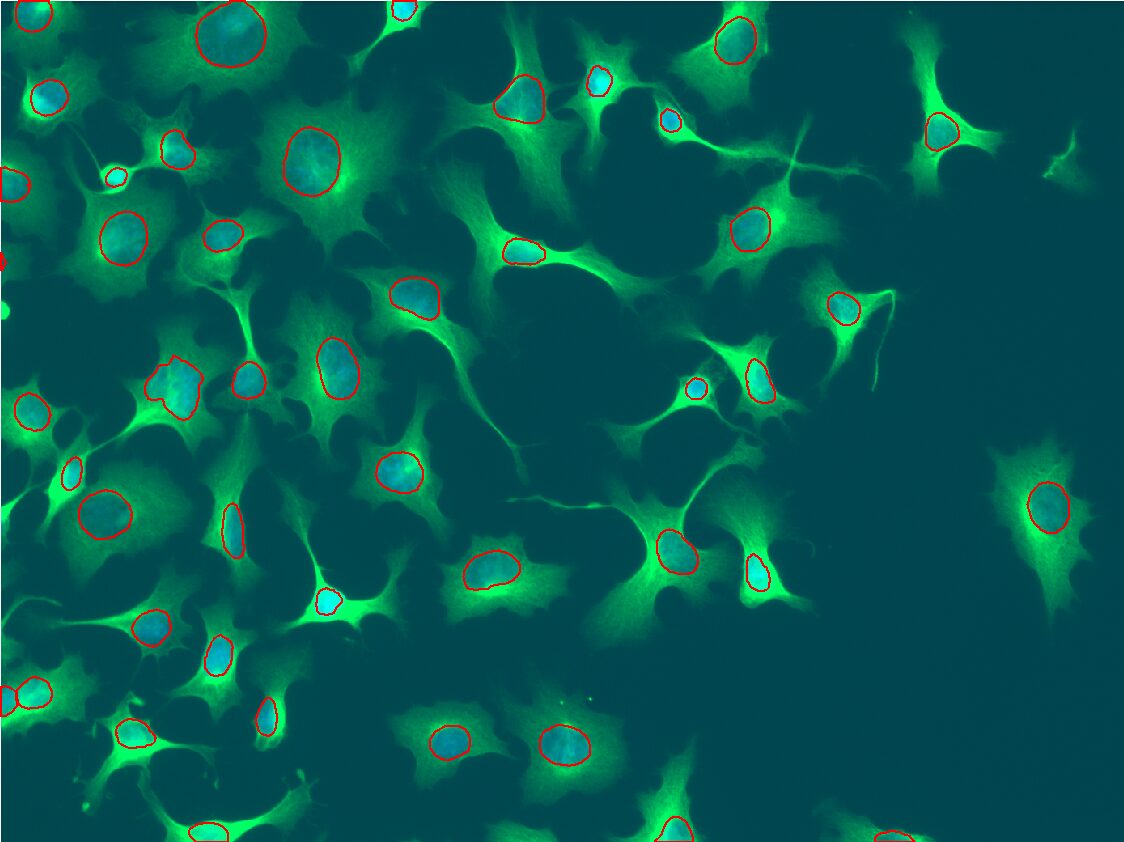
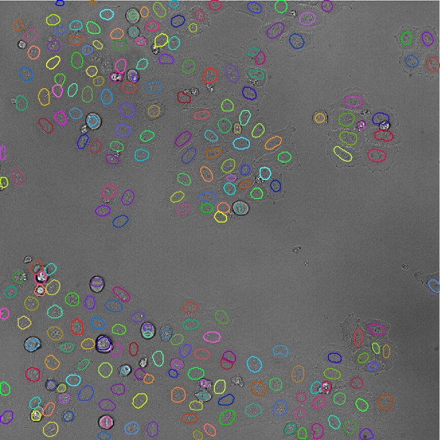
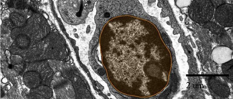
- All
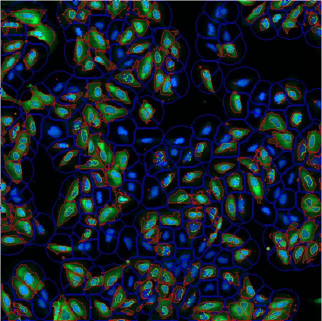
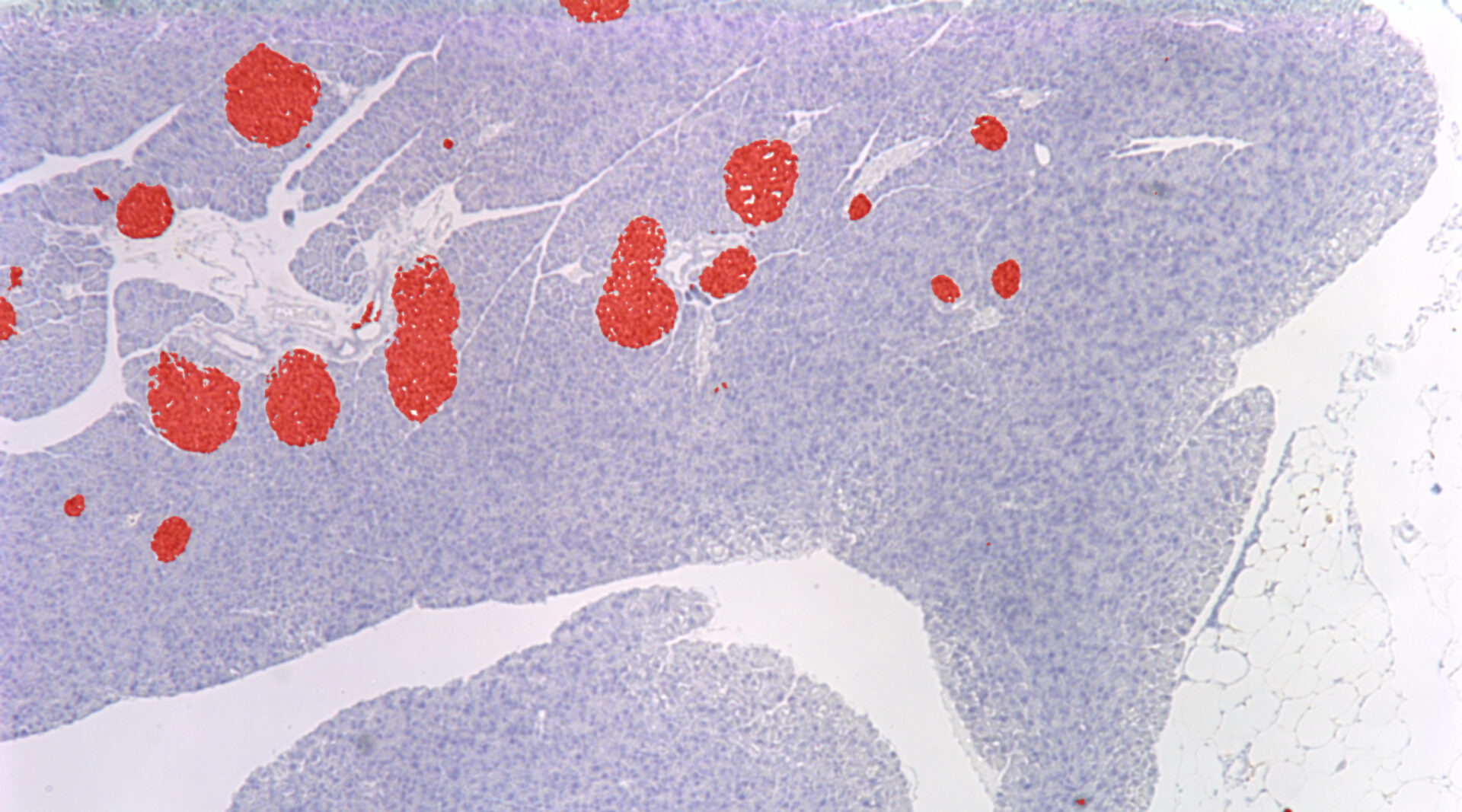
- Biomedical
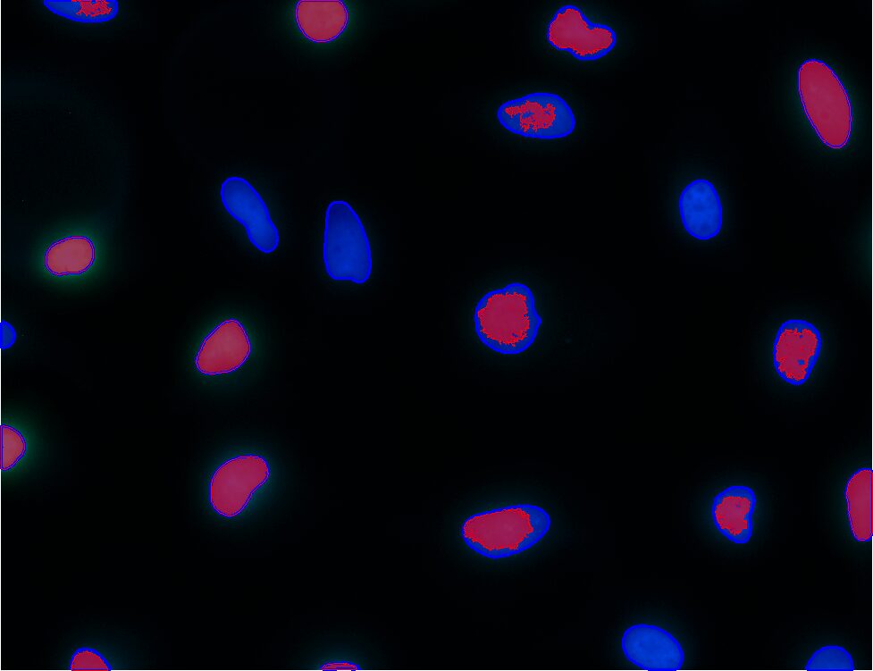
- Cancer Research
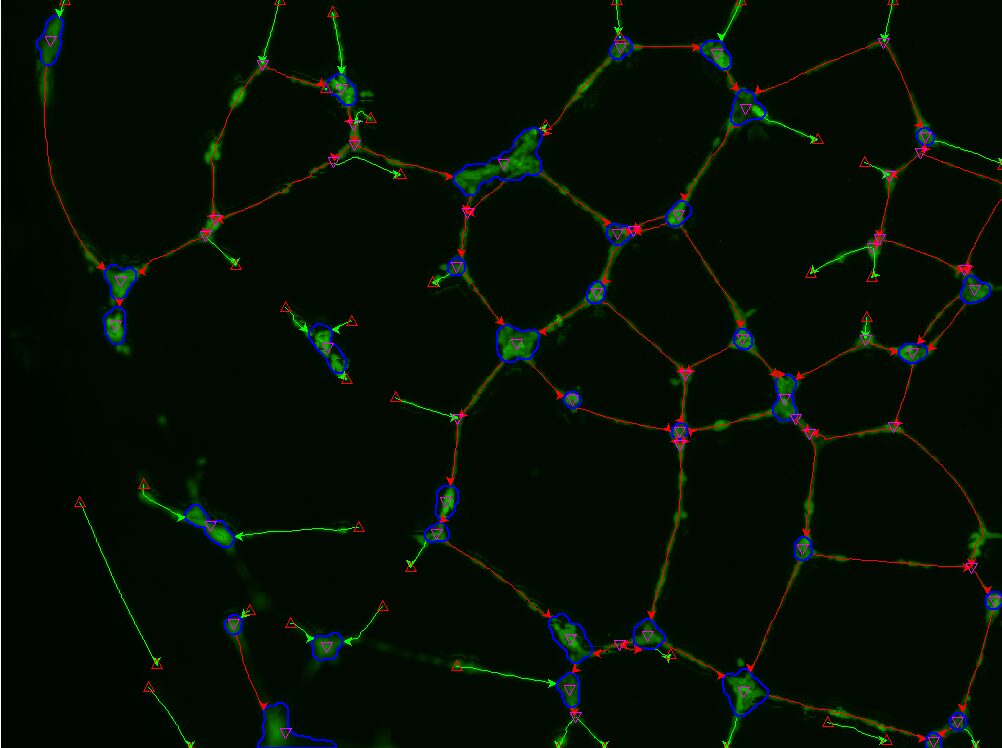
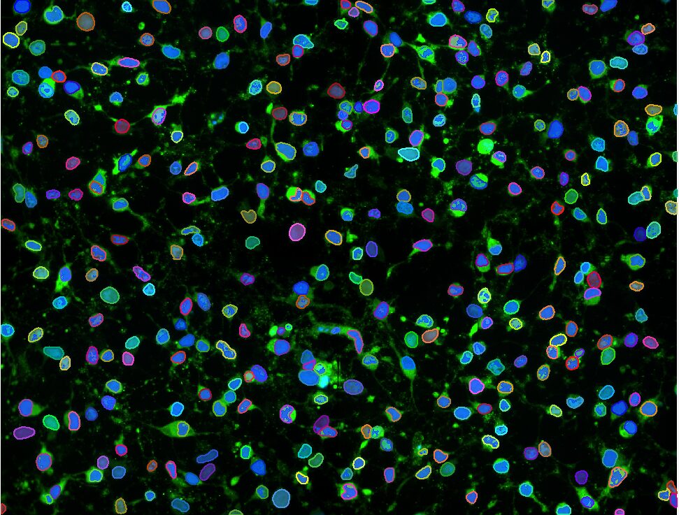
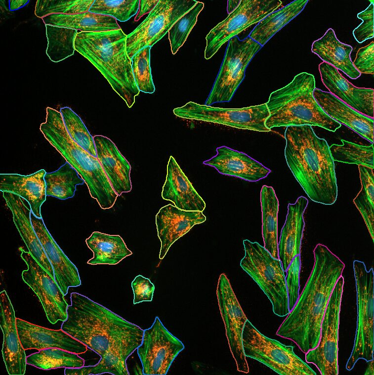
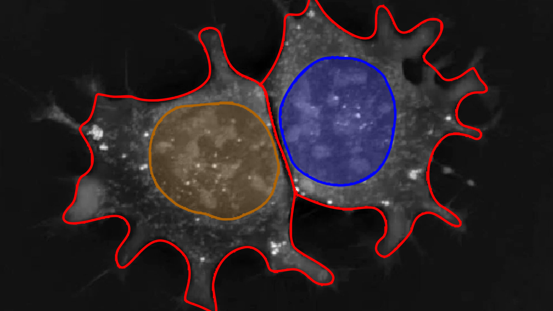
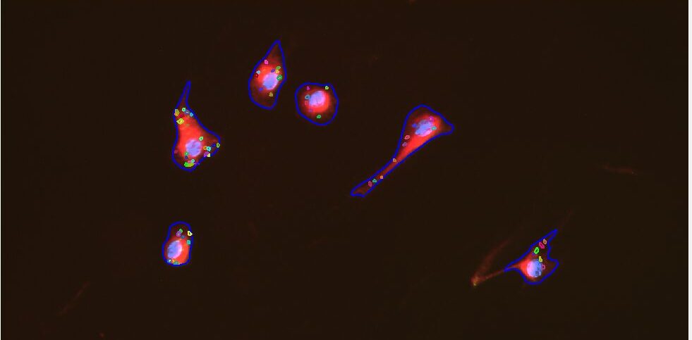
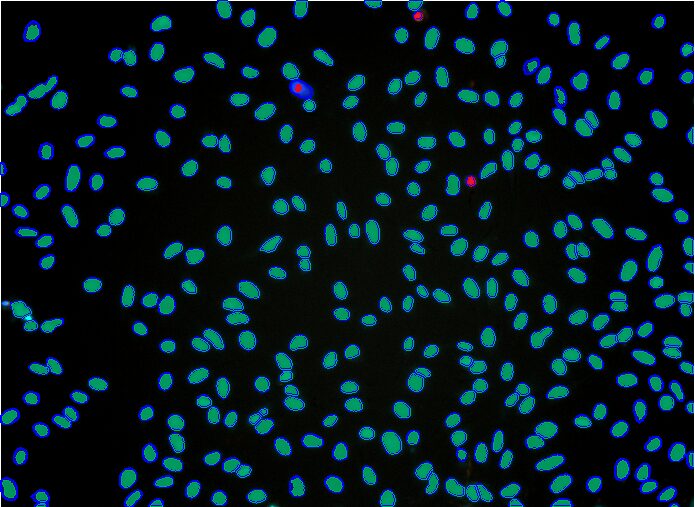
- Cell Biology
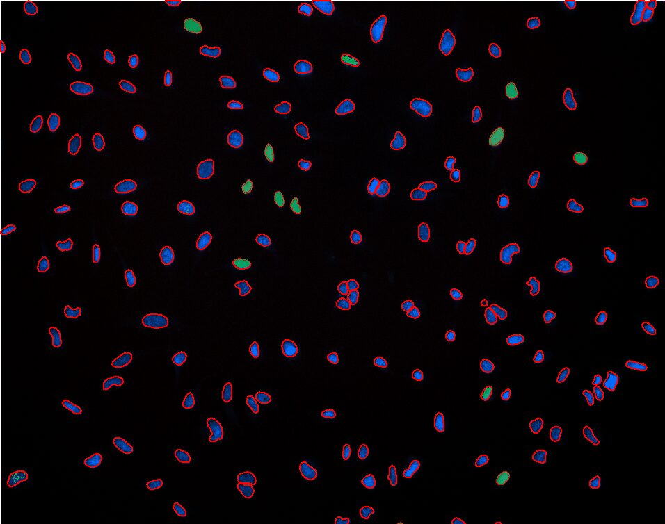
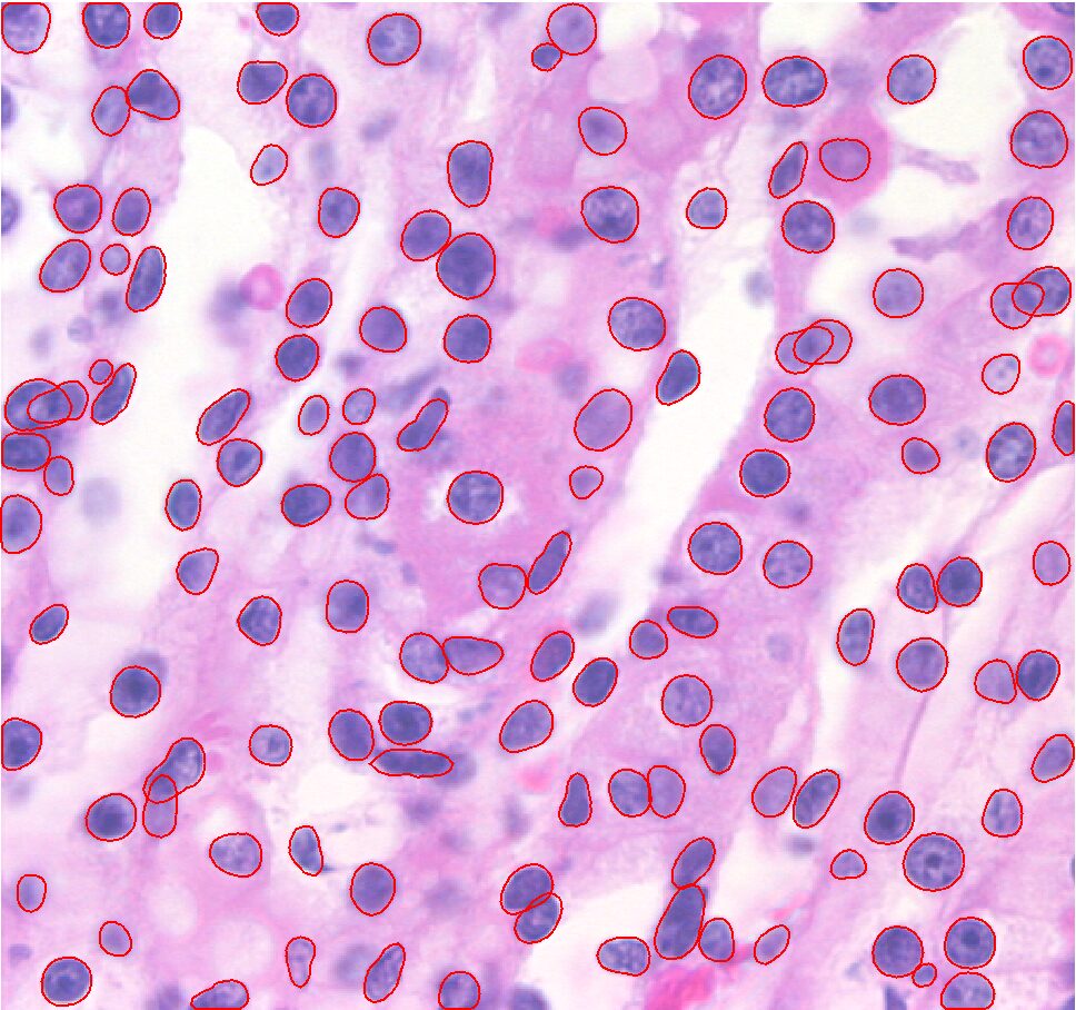
- Histology/Pathology
- New
- Protocol-Guided
- AI-Powered
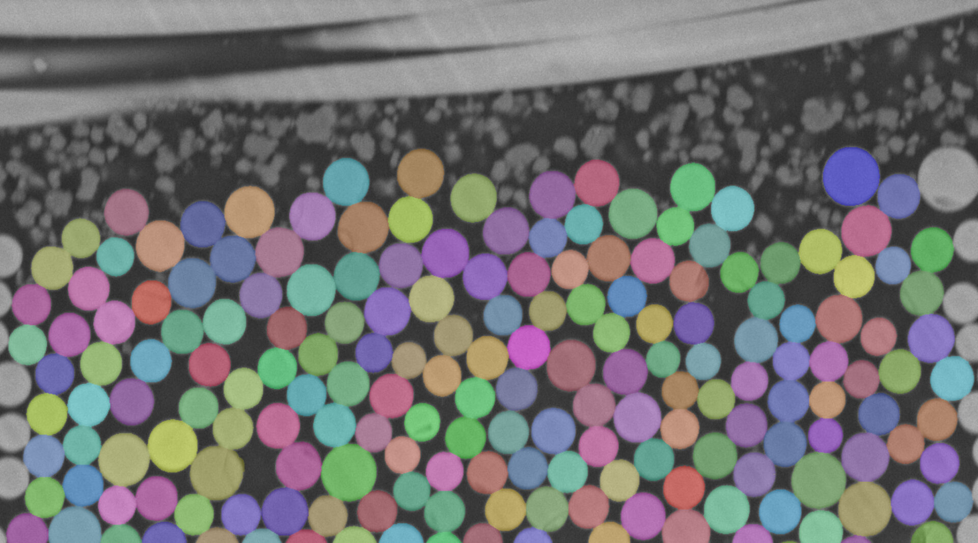
- All
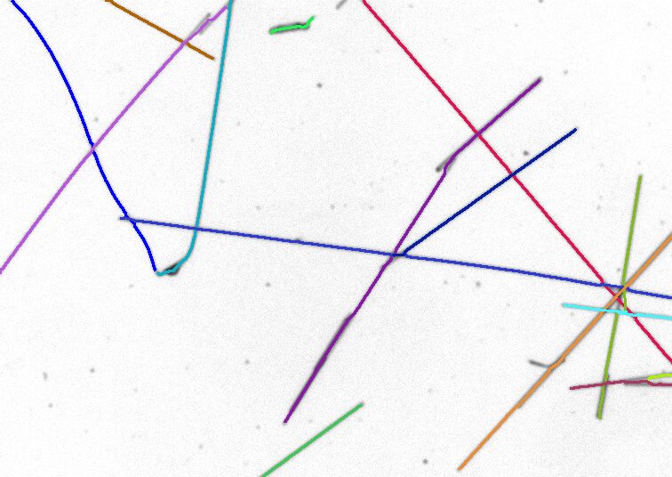
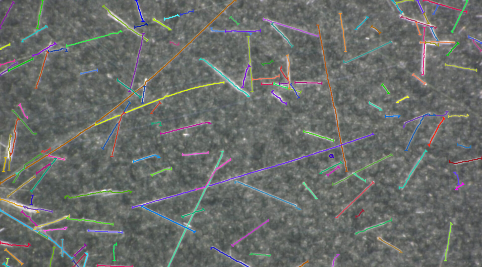
- Area Fraction
- Ceramics & Polymers
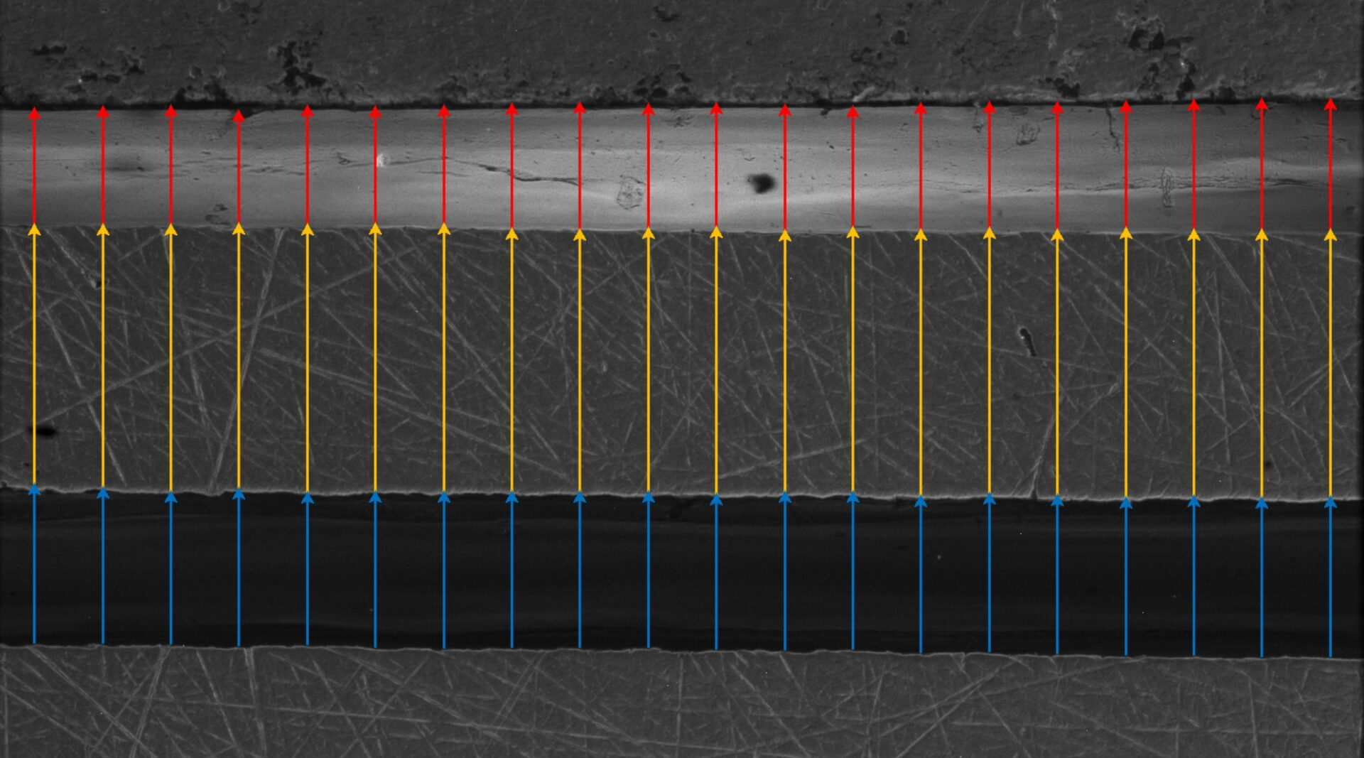
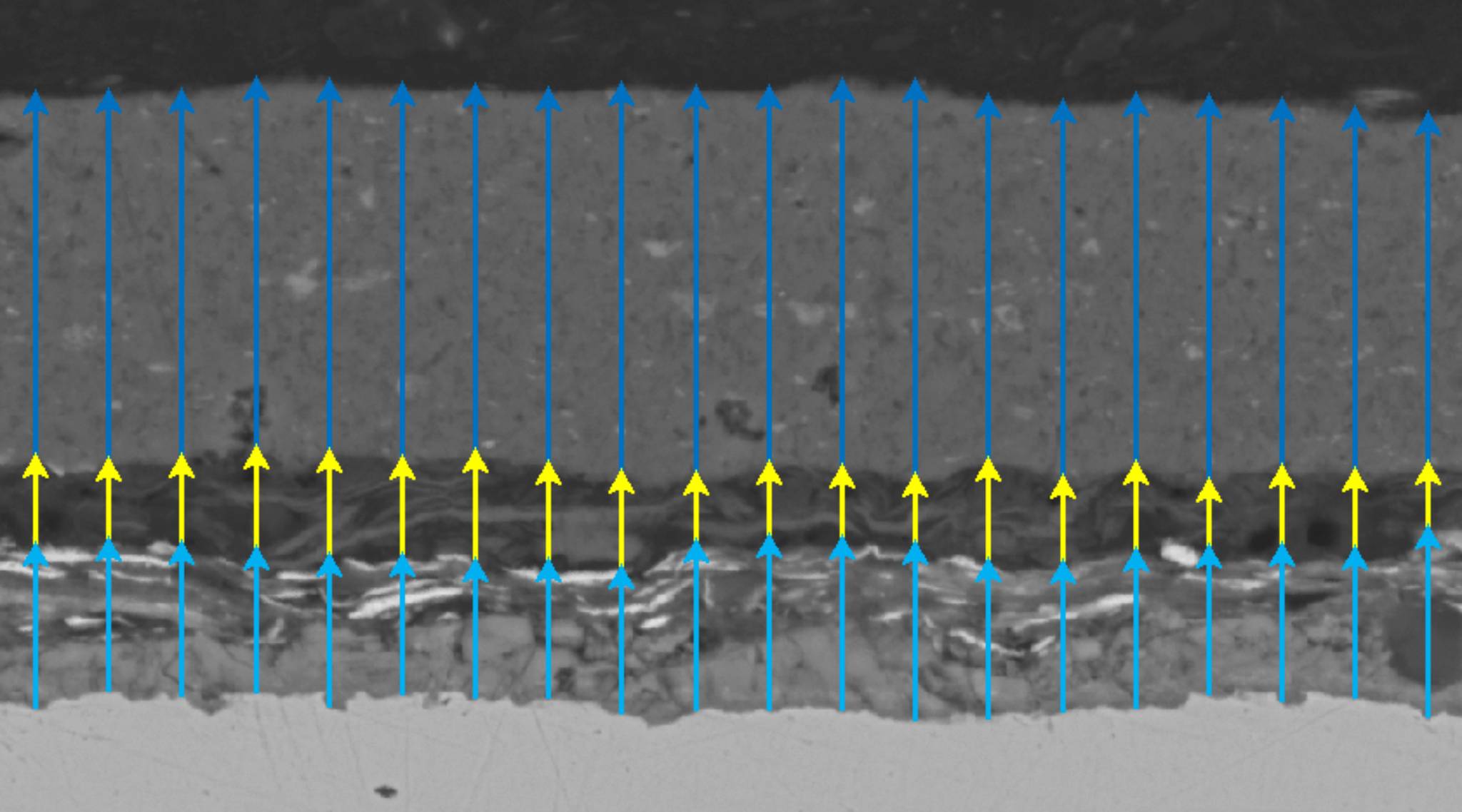
- Coatings & Layers
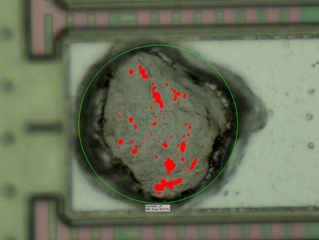
- Electronics QA
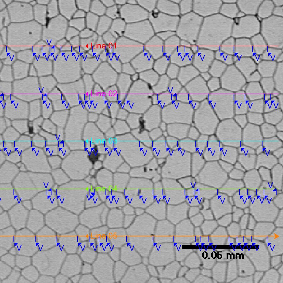
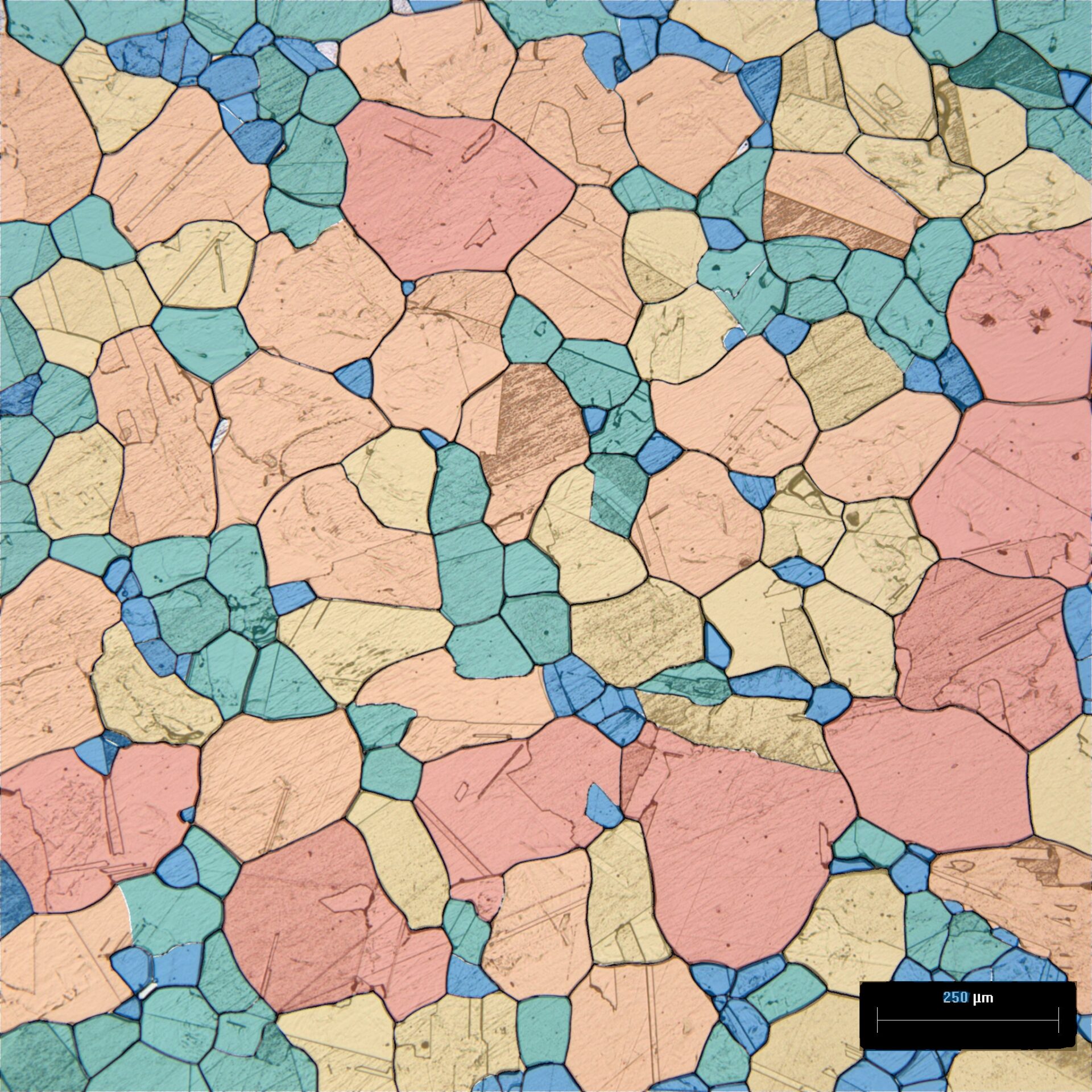
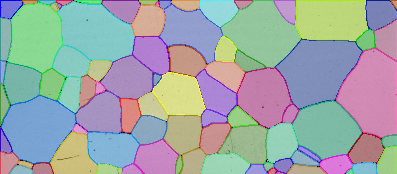
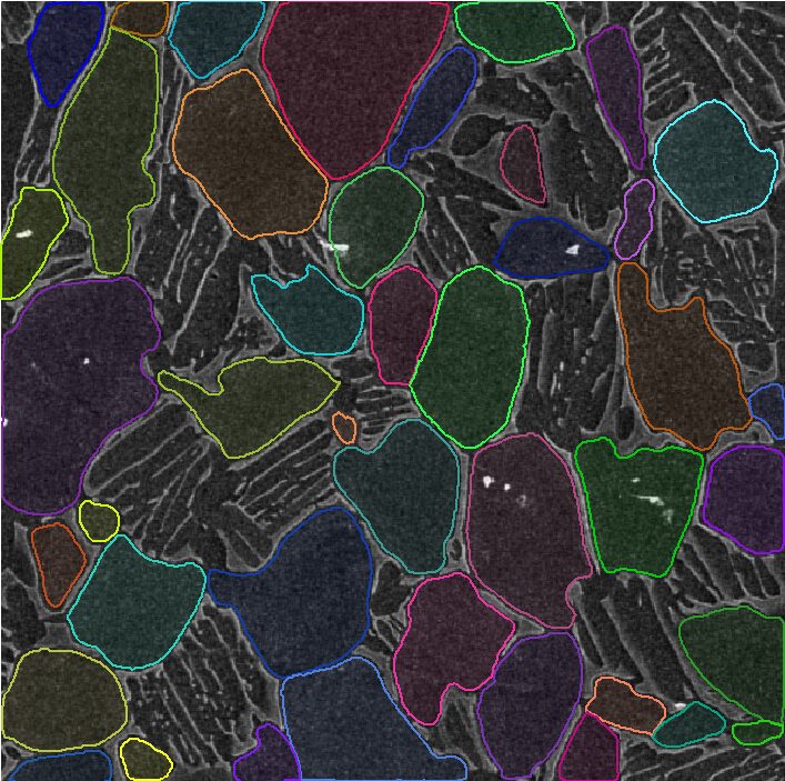
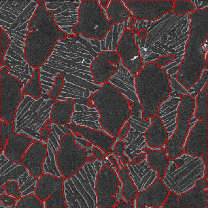
- Grains
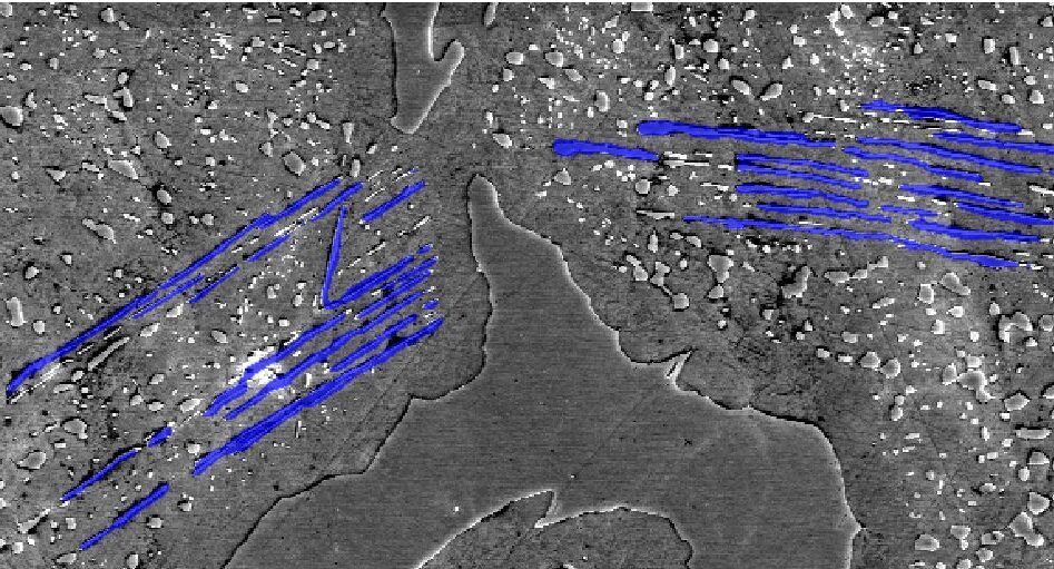
- Metallurgy
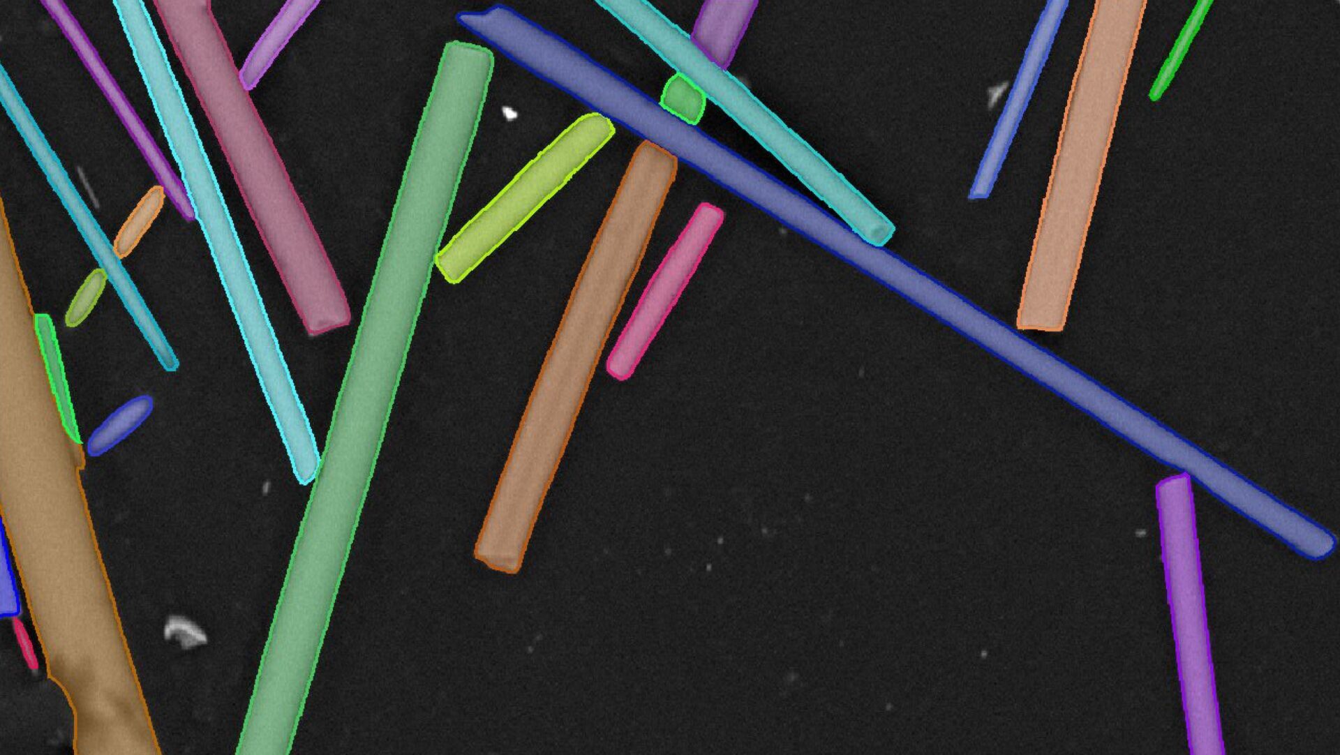
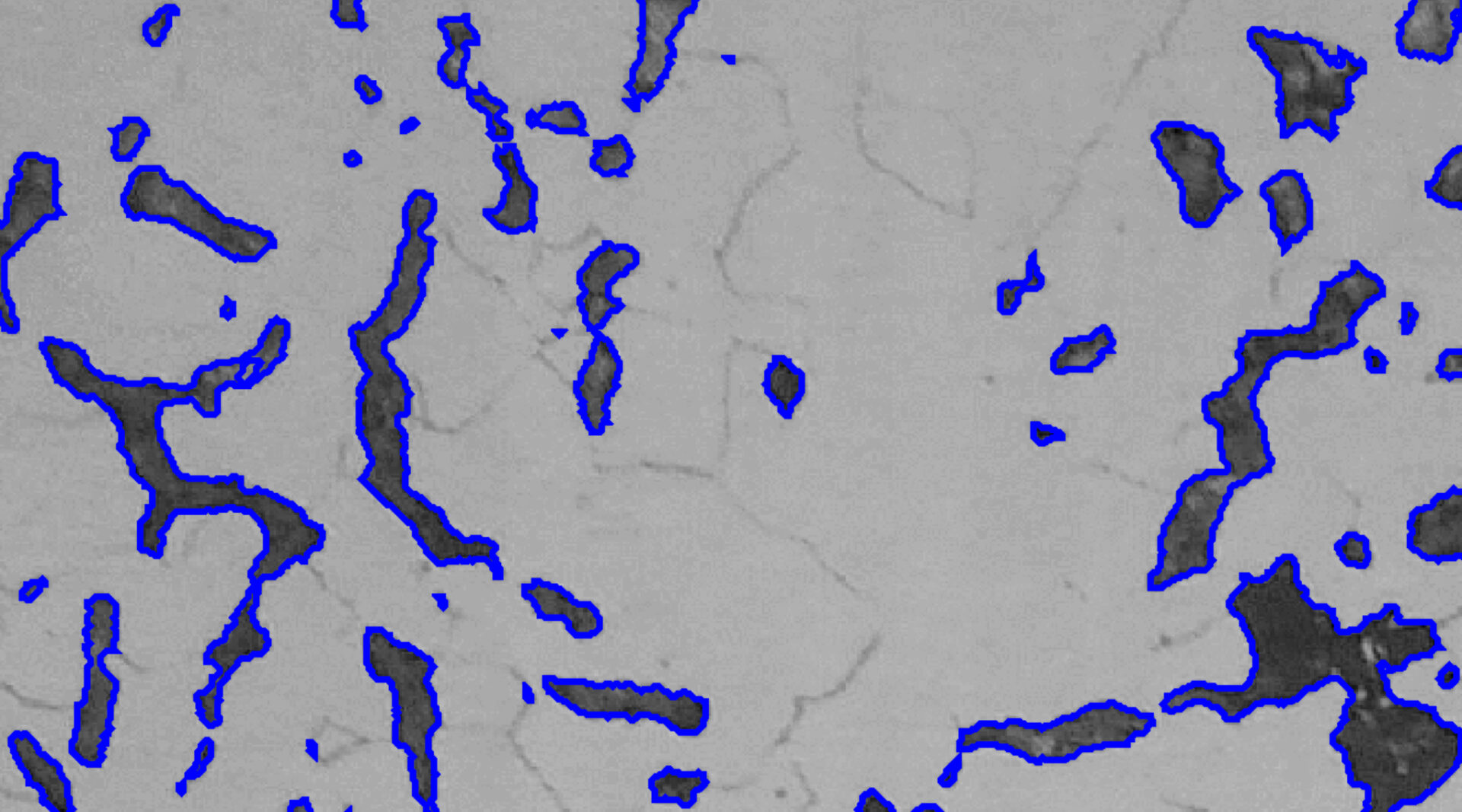
- Particles & Pores
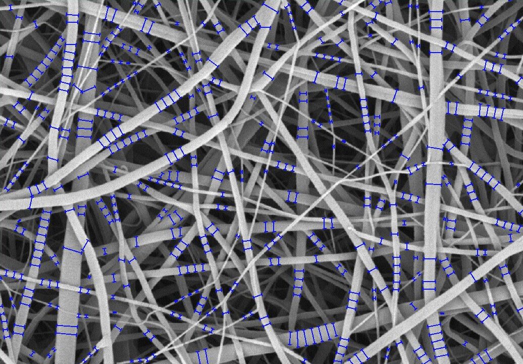
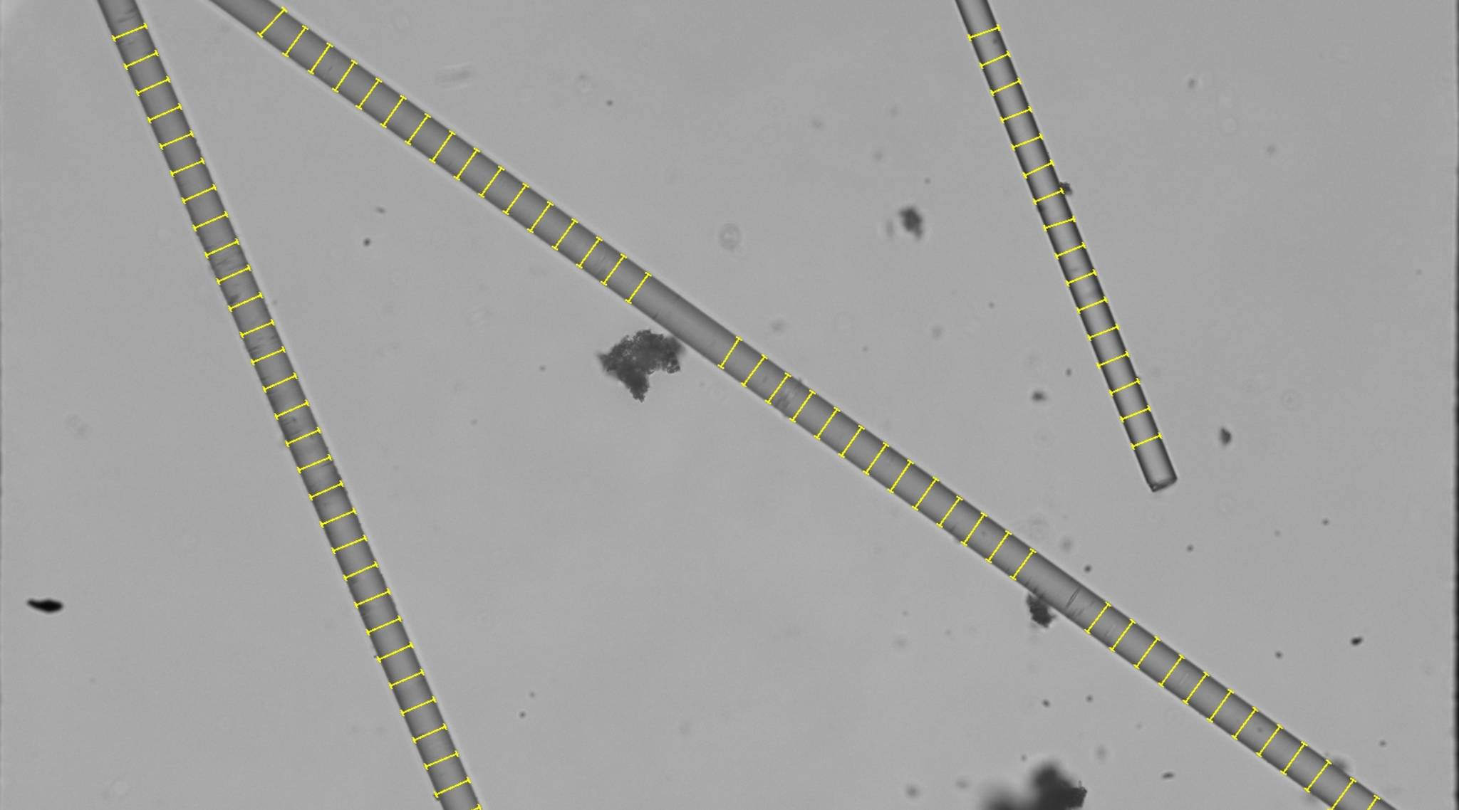
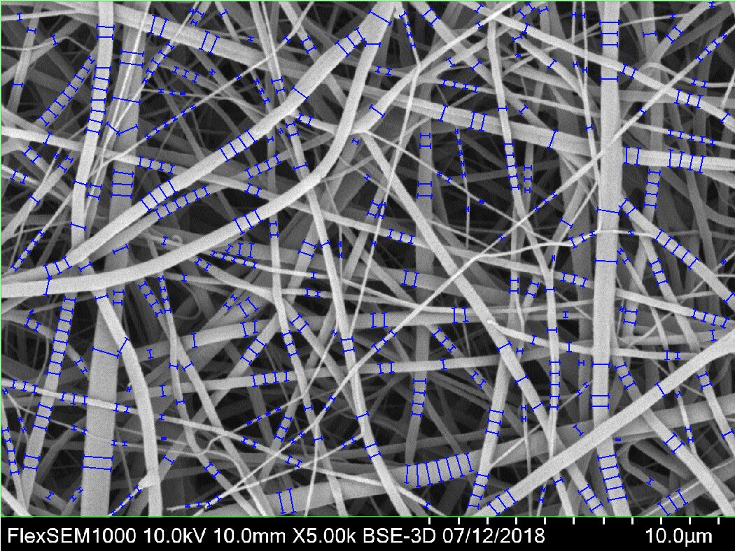
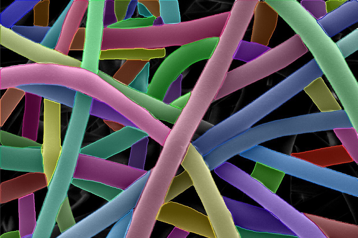
- Textiles & Fibers
- New
- Protocol-Guided
- AI-Powered